ECG interpretation has long been a guilty pleasure of mine. One of the first books I purchased as a medical student was the ‘ECG Made Easy’. After reading that book for the first time ECG interpretation seemed far from easy and I didn’t develop a strong grasp of the subject until many years later.
Whilst working as an A&E SHO many years ago I missed a fairly simple ECG diagnosis, which was fortunately picked up by another doctor that I worked with. This incident highlighted an educational need and my obsession with the ECG has remained ever since. I still look at ECGs on an almost daily basis and it never ceases to amaze me that such a ‘simple’ tool can provide such a wealth of information about a patient. We would not have this remarkable diagnostic tool without a remarkable man, Willem Einthoven.
Who was Willem Einthoven?
Willem Einthoven was born on May 21st, 1860, in Semarang, a coastal city on the island of Java, which was then part of the Dutch East Indies (now Indonesia). He was the son of a doctor and army medical officer, Jacob Einthoven, who had been posted to the Indies. His mother was Louise de Vigel, the daughter of the then Director of Finance in the Indies.
When Einthoven was six-years-old his father died and a few years later his mother decided to move back home to Holland with Einthoven and his two brothers and three sisters. The family settled in Utrecht and Einthoven would attend the University of Utrecht to study medicine a few years later. Einthoven qualified with a degree in medicine in 1885. The following year he was appointed to the position of Professor of Physiology at the University of Leiden. He was a talented researcher and it was during his time at Leiden that he would develop the string galvanometer, an invention that would change the face of medicine and cardiology to this very day.

Willem Einthoven pictured in 1906
Animal Electricity
Long before Einthoven’s time, it was known that the beating of the heart produced electrical currents, but there was no instrument available to measure these currents in an acceptable and non-invasive way.
The long road to the development of the electrocardiogram began with the work of William Gilbert, who was Queen Elizabeth I’s personal physician and President of the College of Physicians (before its Royal Charter). Gilbert published the famous paper ‘De Magnete’ in 1600, his findings suggested that ‘magnetism’ and ‘static electricity’ were separate entities. It was in this paper that he introduced the term ‘electrica’ for insulators that hold static electricity. 46 years later the famous polymath Sir Thomas Browne would refine this term and define ‘electricity’ as “the power to attract straws or light bodies, and convert the needle freely placed”.
The first person to make the observation that electricity existed and played a role in the bodies of animals was the British scientist John Walsh. He managed to obtain a visible spark from an electric eel. He used thin strips of tin foil and demonstrated this technique to many colleagues and visitors in 1773.
Just over a decade later the Italian Anatomist Luigi Galvani made one of the most important observations in understanding the physiological role of electricity when he noticed the twitching of the muscles of a dissected frog’s leg when touched with a metal scalpel. On 20th September 1786 he wrote:
“I had dissected and prepared a frog in the usual way and while I was attending to something else I laid it on a table on which stood an electrical machine at some distance from its conductor and separated from it by a considerable space. Now when one of the persons present touched accidentally and lightly the inner crural nerves of the frog with the point of a scalpel, all the muscles of the legs seemed to contract again and again as if they were affected by powerful cramps.”
Galvani coined the term ‘animal electricity’ to describe the force that activated the muscles of his specimens. The phenomenon was later called ‘galvanism’ after Galvani but today the study of galvanic effects in biology is better known as electrophysiology. Galvani’s name is also given to the ‘galvanometer’ an instrument for measuring and recording electricity, which is integral to the modern electrocardiogram.
The galvanometer was used by the Italian Physics Professor Leopoldo Nobili to detect the flow of an electric current in the body of a frog from the muscles to the spinal cord in 1826. A student of Nobili, Carlo Matteucci, would later observe in 1838 that an electric current accompanies each heartbeat in a frog.
This work of Matteuci would, in turn, influence the German physiologist Emil du Bois-Raymond, who attempted to duplicate his work and ended up discovering the nerve’s ‘action potential’ in 1843. He detected a small voltage potential present in resting muscle and noted that this diminished with contraction of the muscle. To perform this experiment he created an incredibly sensitive galvanometer that needs over 5 km of wire. This galvanometer would later be improved upon by William Thompson (Lord Kelvin) when he created the ‘Thompson Siphon Recorder’ in 1867.
The Road to the Electrocardiogram
It is thought that the first human electrocardiogram was recorded by the Scottish electrical engineer Alexander Muirhead in 1870. He is said to have used a Thompson Siphon Recorder to do so at St. Bartholomew’s Hospital, but this is somewhat disputed as the work was never published.
The British physiologists John Burden Sanderson and Frederick Page would make great strides in the advancement of the understanding of the electrical activity of the heart in the late 1800’s. In 1878 they used an invention called the capillary electrometer to directly record the electrical current within the heart and were the first to note that it consisted of two phases, which would later be recognised as the QRS complex and the T wave.
The first published human electrocardiogram was recorded by the British physiologist Augustus Waller of St. Mary’s Medical School in London. This was also recorded using a capillary electrometer and was performed on a laboratory technician called Thomas Goswell.
Enter Willem Einthoven
Up until this point, the recordings of the heart’s electrical activity were crude and the measuring devices cumbersome. Einthoven witnessed a demonstration by Waller at the First International Congress of Physiology in Basle, Switzerland, in 1889. Waller demonstrated using his pet dog, Jimmy, who apparently sat patiently with his paws in glass jars of saline.

Augustus Waller in his lab and his dog “Jimmy” with his paws in glass jars of saline, image sourced from Wikipedia
Courtesy of Wellcome images CC BY-SA 4.0
Inspired by this demonstration Einthoven made considerable improvements to the recording of cardiac electrical activity with the capillary electrometer and he introduced the term ‘electrocardiogram’ at a meeting of the Dutch Medical Association in 1893. Two years later in 1895 Einthoven managed to distinguish five deflections using an improved electrometer and a correction formula. He named these deflections P, Q, R, S, and T using a mathematical convention dating from Descartes. N has other meanings in mathematics and O is used for the origin of the Cartesian coordinates. P was simply the next available letter and the nomenclature has stood to this day.
Einthoven’s big breakthrough, however, came with his invention of a new galvanometer in 1901. This version of the galvanometer, which he named the ‘string galvanometer’ used fine quartz string coated in silver and weighed approximately 600 lbs. A year later Einthoven published the first electrocardiogram recorded on his string galvanometer and within a year the production of the worlds first commercial ‘ECG machine’ was underway.

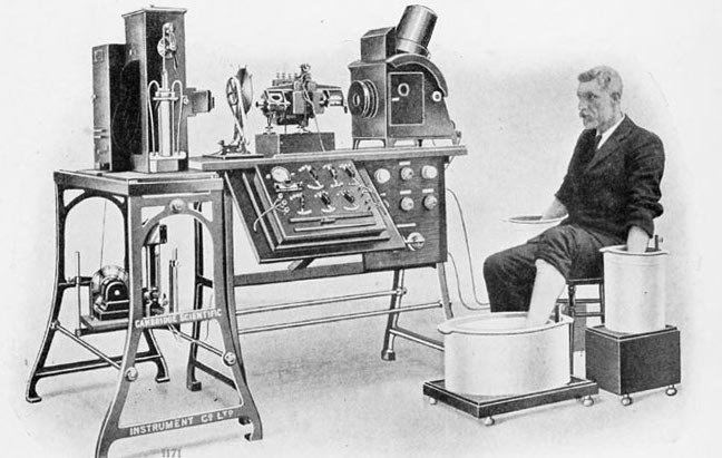
An early ECG machine (circa. 1911)
Einthoven’s Legacy
Following Einthoven’s work, the use of commercial ECG machines became increasingly commonplace. Numerous advances in both recording and interpretation of ECGs would occur over the years that followed. Einthoven received the Nobel Prize in Medicine for inventing the first practical system of electrocardiography used in medical diagnosis in 1924.
The work of Einthoven and the many pioneers of science that went before him have left a remarkable legacy to medicine. The ECG remains one of the most useful investigations available to us in medicine and each and every hospital produces literally hundreds of recordings now on a daily basis. These ECG recordings have resulted in the saving of countless lives due to cardiac problems and will no doubt continue to do so long into the future.


Recent Comments